Introduction to Darkfield Microscopy
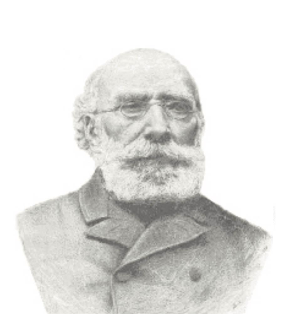
DR. ANTOINE BÉCHAMP,
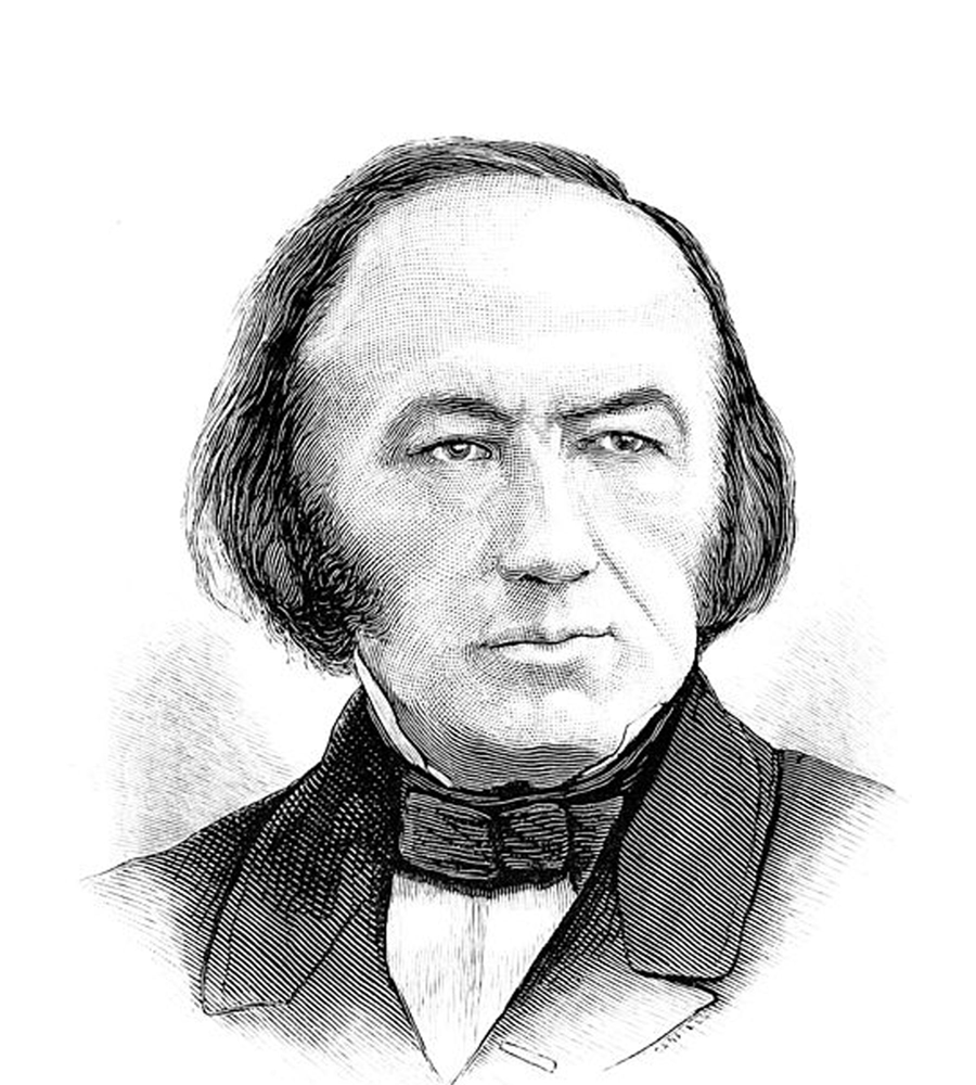
DR. CLAUDE BERNARD,
|
Darkfield microscopy is a special form of microscopy in which the light beam is split in such a way that the edges of objects in the samples are illuminated so that they appear as silhouettes against a dark background — as opposed to brightfield microscopy which allows the examination of specimens against an illuminated field — in which tiny and faint objects are usually washed out. However, these objects can be seen in darkfield and often provide clues as the problems in the sample being studied. The two perspectives can be compared to day and night. In the day time, we see the sun. Because of its brightness, we do not see the stars. At night, we do not see the colors we see in daylight, but we see the stars, millions of them. Stars are there in the day, but we can rarely see them except when rising or setting. In a way, it is fair to say that darkfield viewing is similar to studying the night sky — and what we see in darkfield is similar to the night because mysteries are often revealed. One sometimes has the feeling of looking deep into inner space. Historically, darkfield microscopy has often been associated with two critical ideas, usually attributed to Dr. Claude Bernard (1813-1878) and Dr. Antoine Béchamp, (1816-1908):
Bernard held the first chair in physiology at the Sorbonne and was highly regarded as a true scientist. He emphasized the need to accept facts, even if they are opposed by prevailing opinions. Béchamp is perhaps best known for losing the debate with Louis Pasteur, thus leading the war on germs that characterized 20th century medicine to the present times. From the perspective of the terrain theorists, the alternative would be to cultivate the internal conditions that support health and immunity. The insights of these illustrious Frenchmen influenced the work of such innovative thinkers as Prof. Gunther Enderlein, Dr. Royal Raymond Rife, Dr. Wilhelm Reich, Gaston Naessens, Dr. Virginia Livingston-Wheeler, Dr. Alan Cantwell, Prof. Erik Enby, and, of course, many others. The method of observation is just that: a way of collecting impressions; and it is important to point out that a sample of blood is viewed while living so there is movement, change, reaction, organization, and much more than can be observed when using a fixative or damaging the sample by smearing it. The viewing options are facilitated by very minor modifications of a basic light microscope. However, the conclusions that are drawn may or may not conform to prevailing views. Therefore, it is best to keep an open mind and use discernment before accepting or rejecting one view or another. Because darkfield is not generally taught in medical schools, different techniques and protocols tend to emerge based on the interests and training of the practitioners. In my case, with a background in anthropology, including ethnobotany, I may be predisposed to observe so I refer to myself as a blood behaviorist. By giving herbs, the responses can be assessed and protocols can be tweaked until the plasma is healthy and both the erythrocytes and white blood cells have the perfect milieu for supporting the health of the individual. It is worth reiterating that there is nothing inherent in the configuration of the microscope that predisposes one to believe in pleomorphism over monomorphism. If the observations support one conclusion over the other, then one can take a position in this ongoing debate. My goal is to help people to identify and correct whatever issues are found. The course that I teach will begin with how to choose the best scope for the budget available. Once the microscope is delivered, the assembly, correct operation and maintenance of the scope, and method of sampling, focusing, photographing, and archiving images will follow. This part goes relatively quickly so people are up and running in a day or two. The interpretation of the issues and mastery of the protocols is truly a major study and is expected to span at least two years. This website is a membership site. There are only a few pages that guests can view. Everything else is handled very securely to protect the privacy of the patients, but the tools for uploading histories, photomicrographs, videos, protocols, and updates are all built into the site. There is a forum for discussion, downloadable material, charts that can be used for reference or decorating offices, and access to the herbs that can be used for correcting whatever problems are found. First posted by Dr. Ingrid Naiman on 28 February 2007 and updated 23 September 2014 and 23 May 2022 |